


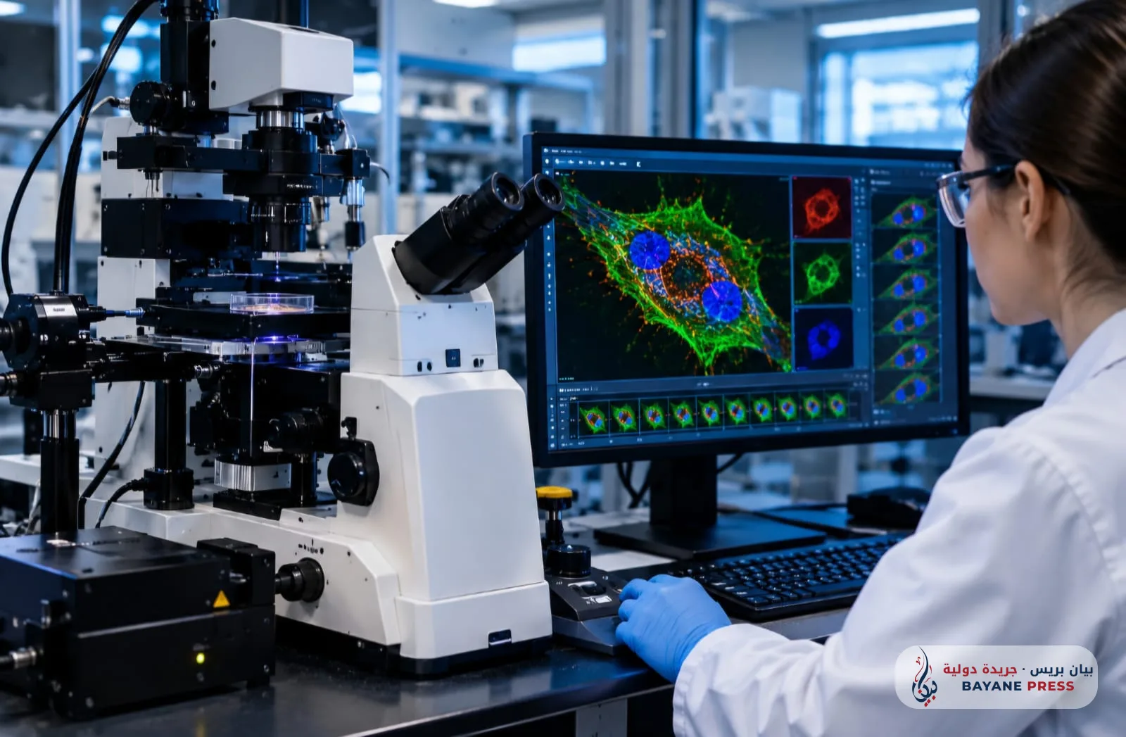
Researchers in the bioengineering laboratories at Stanford University, in cooperation with the Max Planck Institute for the Science of Light, revealed on October 16, 2026, the development of an advanced generation of the super-interferometric scanning microscope, technically known as the enhanced iSCAT system.
The paper published in the journal Nature Methods demonstrated the system's ability to image precise biological processes inside living cells with ultra-high quality and without the use of any chemical labeling media or fluorescent dyes, overcoming the major obstacle in microscopy represented by phototoxicity and signal fading over time.
The new microscope relies on measuring the optical interference between light reflected from a reference glass interface and the extremely faint light scattered by individual biological molecules inside the cell. The team developed optical algorithms and ultra-low intensity laser illumination at microwatt levels, allowing the image capture speed to be raised to more than 1,000 frames per second with a spatial resolution approaching 2 nanometers. This enabled the documentation of the movement of motor proteins such as kinesin and myosin during the transport of vesicles across microtubules within the cytoplasm, and the tracking of receptor diffusion across the cell membrane with extreme precision.
This innovation represents a crucial tool for accelerating pharmacological research and oncology treatments, as it allows scientists to examine the mechanisms by which vital drug molecules penetrate and cross cell membranes in their natural physiological environment without causing distortion to the cell structure, while reducing laboratory preparation costs and accelerating preliminary clinical trials.




Comments (0)
No comments yet. Be the first to react!